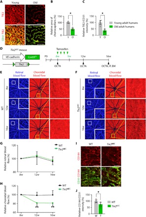
Fig. 1. Tie2 deletion in adult ECs leads to damage and loss of choriocapillaris.

(A to C) Images and comparisons of density and TIE2 intensity of CD31+ choriocapillaris in healthy young adult (20 to 36 years old; Y) and old adult (65 to 84 years old; O) human subjects. Error bars represent means ± SD. Each group, n = 5. *P < 0.05 versus Young by Mann-Whitney U test. Scale bars, 75 μm. (D) Diagram of schedule for EC-specific depletion of Tie2 in 8-week-old mice and intravital OCTA at 8, 12, and 16 weeks (w) of age using Tie2iΔEC mice. (E and F) En face OCT angiograms showing retinal blood flow (left) and choroidal blood flow (middle) acquired by longitudinal OCTA imaging of eyes. Each area marked by a white box is magnified in the corner. Each area marked by a yellow box is magnified on the right panel. (G and H) Temporal changes in relative retinal (G) and choroidal (H) blood flow. Error bars represent means ± SD. Each group, n = 10. **P < 0.005 versus WT by unpaired Student’s t test. (I and J) Images and comparison of CD144 intensity in CD31+ choriocapillaris. Error bars represent means ± SD. Each group, n = 5. *P < 0.05 versus WT by Mann-Whitney U test. Scale bars, 20 μm.
