
Fig. 1. Vps4 is required for axonal integrity and its OE delays WD.
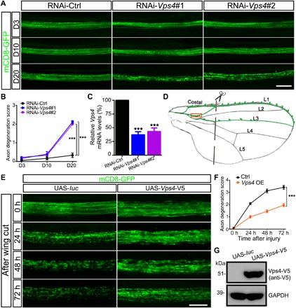
(A and B) Representative images of the wing axons labeled by mCD8-GFP of control (RNAi-Ctrl) or RNAi-Vps4 flies at indicated ages. Axonal degeneration scores are evaluated as described in fig. S1 and quantified in (B). Data shown are means ± SEM; n = 7 to 10 wings per time point per genotype; ***P < 0.001; two-way analysis of variance (ANOVA). D3, day 3; D10, day 10; D20, day 20. (C) The KD efficiency of the RNAi-Vps4 lines is analyzed by quantitative polymerase chain reaction (qPCR) and normalized to actin. Means ± SEM; n = 3; ***P < 0.001; Student’s t test. (D) A schematic drawing of the Drosophila wing, highlighting the neuronal soma and axons in the costal, L1, and L3 wing veins. A pair of scissors indicates the injury site, which completely severed all axons of the L1 nerve, and the boxed area is imaged in (E). (E and F) Representative images (E) and quantification (F) of mCD8-GFP–labeled wing axons of the control (UAS-luc) or Vps4 OE flies (UAS-Vps4-V5) at indicated time points after axotomy. UAS, upstream activation sequence. Means ± SEM; n = 10 to 12 wings per time point per genotype; ***P < 0.001; two-way ANOVA. (G) Western blotting analysis confirming the expression Vps4-V5 in the transgenic flies. Scale bars, 20 μm (A) and 10 μm (E). GAPDH, glyceraldehyde-3-phosphate dehydrogenase.