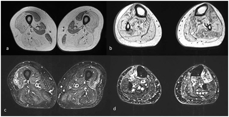
Figure 3.
Muscle MRI axial T1 (a,b) and T2 STIR images (c,d) of the lower limbs shows mainly complete fatty substitution of both anterior and posterior thigh muscles, with selective sparing of the medial ones, specifically rectus, gracilis, sartorius, and adductor magnus (a). Diffuse adipose involvement also of the leg muscles, less pronounced at the level of the soleus, and tibialis anterior muscles (b). Inhomogeneous STIR hyperintensity was also associated, mainly in the quadriceps at the level of the thigh and in the tibialis anterior at the level of the leg (c,d).

