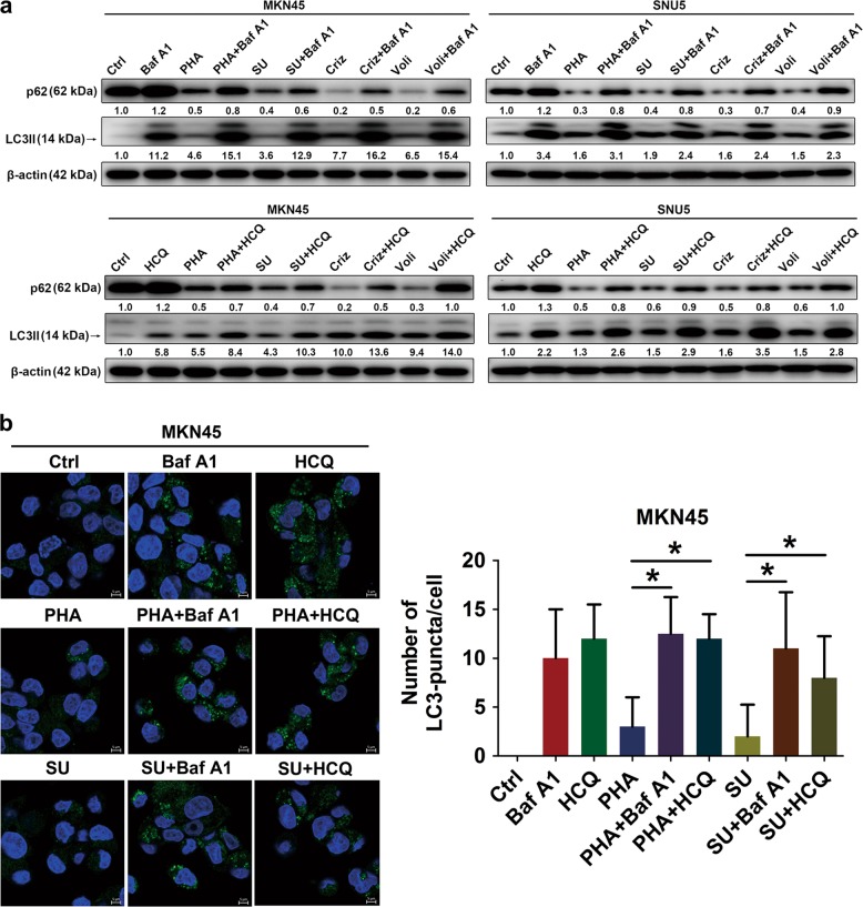
Fig. 2. Met tyrosine kinase inhibitors (Met-TKIs) activated autophagy flux in Met-amplified gastric cancer (GC) cells.
a, b After exposure to Met-TKIs (PHA-665752 (PHA) 200/600 nM, SU11274 (SU) 1/0.8 μM, crizotinib (Criz) 100/50 nM or volitinib (Voli) 10/4 nM for MKN45 and SNU5 cells, respectively) with/without autophagy inhibitors (bafilomycin A1 (Baf A1) 10 nM or hydroxychloroquine (HCQ) 10 μM) for 36 h, cell lysates were immunoblotted for indicated proteins and LC3-positive puncta were counted by confocal immunofluorescence (autophagosomes were green and 4′,6-diamidino-2-phenylindole (DAPI)-stained nuclei were blue). Scale bar, 5 μm. Data are expressed as median with interquartile range. *P < 0.05 by nonparametric Mann–Whitney U-test

