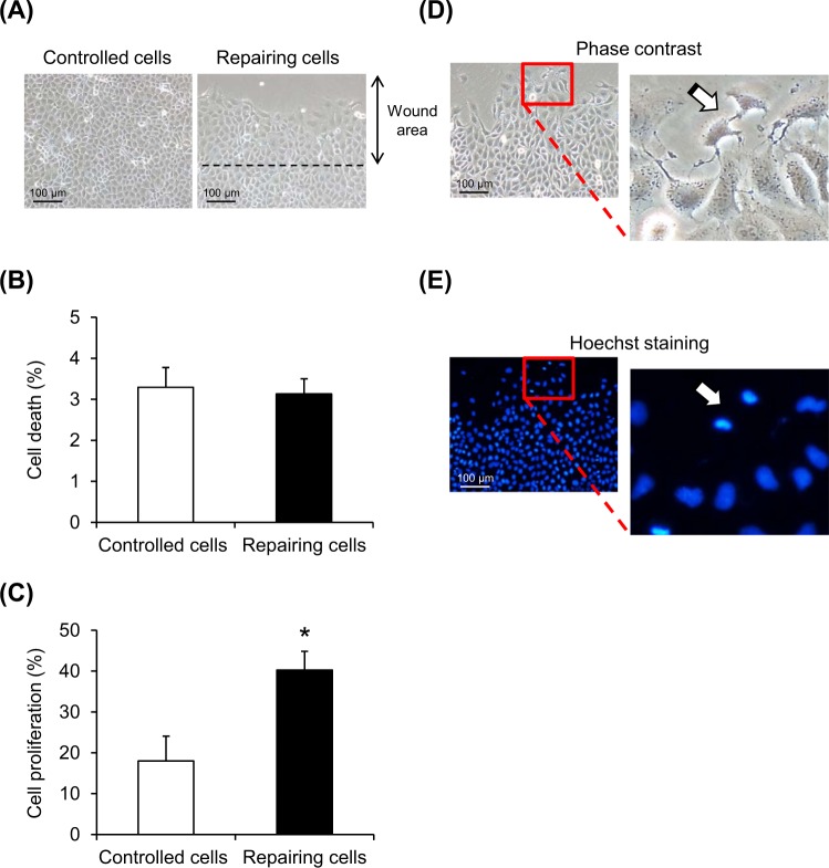
Fig. 2. Enhanced cell proliferation in repairing cells.
At optimal post-scratch time-point (12 h) and crystal-exposure time (30 min), the repairing and controlled cells were subjected to morphological examination (a), cell death assay (b), and cell proliferation assay (c). Phase contrast microscopy (d) and immunofluorescence staining of cellular nuclei (e) were also performed to demonstrate dividing cells. Original magnification =×200 for all panels. Each bar represents mean ± SEM of the data obtained from three independent experiments. *p < 0.05 vs. control

