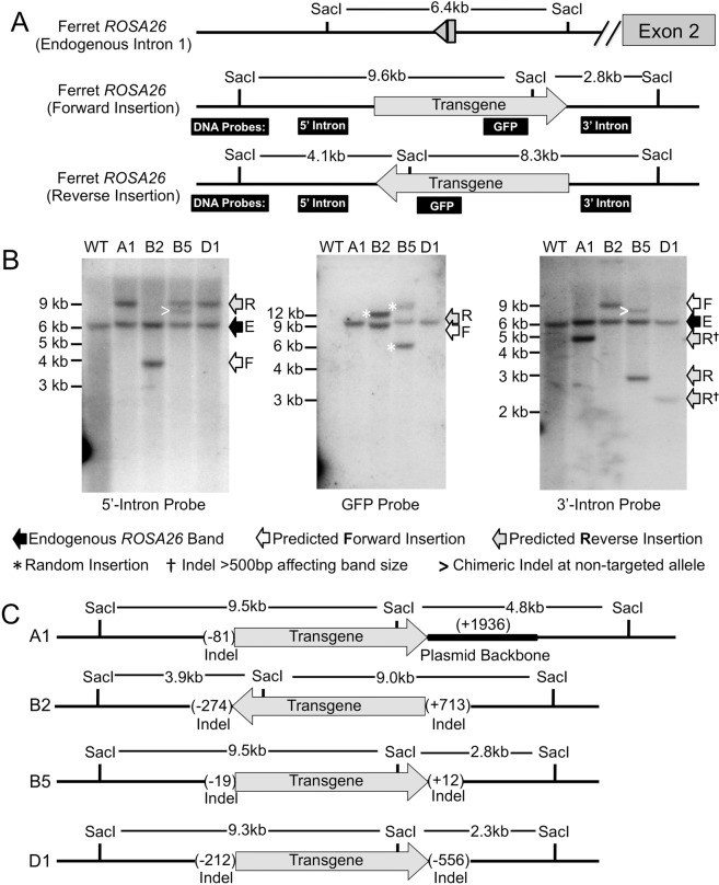
Figure 5.
Southern blot analysis of transgenic founder genomic DNA. (A) Schematic drawing of hybridization probes for the external arms of 3′-intron and 5′-intron and the EGFP transgene, with predicted sizes of Sac1 digested bands for the non-targeted and forward- and reverse-integrated transgenes, respectively. (B) Representative Sac1-restricted Southern blots following hybridization with the indicated probes for the four surviving transgenic ferrets. Arrows to the left of each blot denote bands for the endogenous intact locus (E), forward (F)- and reverse (R)-orientated insertion of the transgene cassette. The extra bands seen in B2 and B5 transgenic ferrets with the EGFP probe (white asterisks) are suggestive of a random integration event in these animals. Other annotations on the blots are marked in the legend at the bottom. (C) Schematic showing the predicted transgene orientation and size of indels at both the 5′ and 3′ junctions of the transgene insertion site for the indicated transgenic animals as determined by results of sequencing and Southern blotting.

