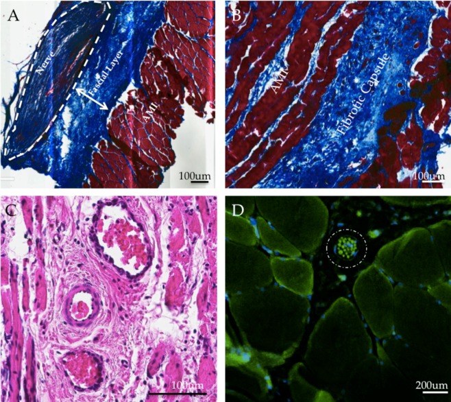
Figure 5.
Histological Results. (A) Well-demarcated partition between the fascial layer and AMI. This representative slice captures a healthy collagen-bound nerve entering the muscle layer and reinnervating the muscle. (Trichrome stain). (B) The fibrotic capsule surrounding the upper layer of the AMI, through which new vessels formed, is comprised of loose, anisotropically oriented collagen. (Trichrome stain). (C) Well-epithelialized angiogenic vessels through scar bed facilitate revascularization. Surrounding musculature appears healthy and mature. (H&E stain). (D) A spindle fiber (circled) is shown containing numerous intrafusal fibers between motor unit groups (s46 stain).

