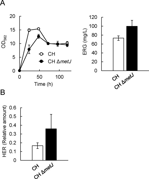
Figure 3.
Impact of metJ gene disruption on ERG and HER productivities. (A) CH pQE1a-egtABCDE and CH ΔmetJ pQE1a-egtABCDE were cultured in SM1 liquid medium. After 6 h cultivation, IPTG and Na2S2O3 were added at concentrations of 0.1 mM and 10 mM respectively. Cell density (left) was estimated from the OD at 562 nm. ERG (right) in the cultured supernatant at 120 h was quantified by Sulfur index analysis described in Methods. (B) WT and ΔmetJ cells harboring pCysHP and pCF1s-egtD were cultured in SM1 liquid medium. After 6 h cultivation, IPTG and Na2S2O3 were added at concentrations of 0.1 mM and 10 mM respectively. HER in the cultured supernatant at 120 h was quantified by LC-MS/MS analysis. Data are presented as mean values with standard errors from three independent experiments.

