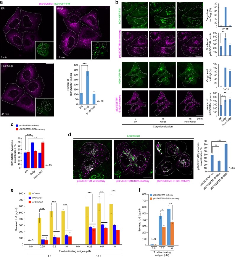
Fig. 7.
The autophagy cargo receptor p62/SQSTM1 is required to sustain protein secretion during lysosome repositioning. a HeLa cells expressing human growth hormone fused to the polymerization/depolymerization FM domain (hGH-GFP-FM) were subjected to the endoplasmic reticulum (ER)-to-Golgi cargo transport assay and then subjected to immuno-staining to detect endogenous p62/SQSTM1. Representative images show p62/SQSTM1 puncta distribution at the indicated times of hGH-GFP-FM localization in the ER, Golgi or post-Golgi (insets) after addition of DD-solubilizer. The graph depicts the quantification of the number of p62/SQSTM1 puncta at the times indicated in the images (n = 50 cells). b HeLa cells expressing hGH-GFP-FM were transfected to co-express p62/SQSTM1-wt-mcherry or the p62/SQSTM1-S182A-mcherry mutant. After 16 h, cells were subjected to the ER-to-Golgi cargo transport assay. Representative images show the localization of hGH-GFP-FM and the distribution of puncta containing either of the p62/SQSTM1-mcherry variant, in conditions as those shown in a. The graphs depicts either the quantification of hGH-GFP-FM in the Golgi complex or the quantification of the number of puncta containing either of the p62/SQSTM1-mcherry variant at the times indicated in the images (n = 15 cells). c HeLa cells transfected to express either of the indicated p62/SQSTM1-mcherry variants were incubated with Lysotracker. Cells were nutrient-starved by incubation for 1 h in Hank’s balanced salt solution. At the indicated times after, images were acquired to calculate the level of co-localization between lysosomes and puncta containing either of the indicated p62/SQSTM1-mcherry variant (n = 15 cells). d HeLa cells were transfected and incubated with Lysotracker as in c, but maintained in regular culture medium. Images were acquired to calculate the level of co-localization in steady-state conditions between lysosomes and puncta containing either of the indicated p62/SQSTM1-mcherry variant. The quantification is shown in the graph at the right of the panel (n = 50 cells). e Mouse LMR7.5 T-lymphocytes were subjected to control silencing (shControl) or silencing of KDELR1 using either of the indicated short hairpin RNA (shRNA). Cells were subjected to an antigen-peptide presentation assay using the indicated concentrations of antigen for the indicated periods of time. To determine T-lymphocyte activation, the level of secreted interleukin-2 (IL-2) was estimated using an enzyme-linked immunosorbent assay (ELISA) assay (n = 3 independent expriments). f Mouse LMR7.5 T-lymphocytes were transfected to express p62/SQSTM1-mcherry-wt or p62/SQSTM1-mcherry-S182A. After transfection, cells were subjected to an antigen-peptide presentation assay and to estimation of T-lymphocyte activation as shown in e (n = 3 independent experiments). Data are means ± SEM. Scale bar, 10 µm. *p < 0.05; **p < 0.01; ***p < 0.001 (Student’s t tests). wt Wild type

