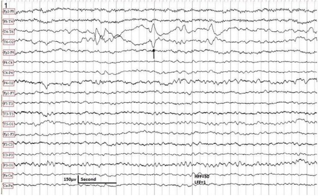
Figure 1.

The EEG sample taken in a 10-year-old male with partial epilepsy controlled by carbamazepine. Patient awake. The EEG shows positive sharp waves in the right posterior temporal region (arrow). Neurological examination was normal. No MRI was available. HFF, high frequency filter; LFF, low frequency filter; EEG, electroencephalography; MRI, magnetic resonance imaging.
