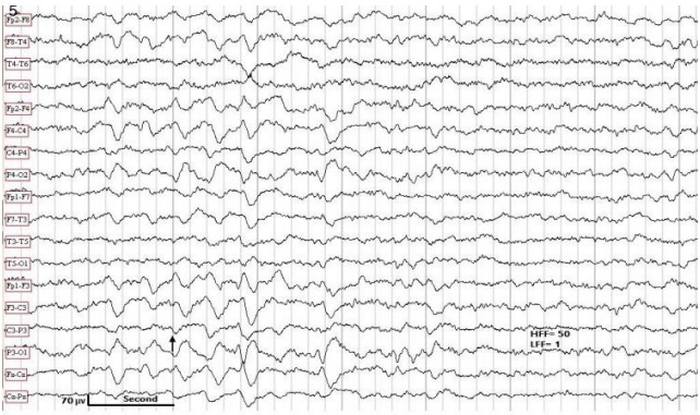
Figure 5.

EEG obtained in a 7-year-old female with generalized convulsive seizures. Patient awake. The EEG shows a burst of generalized but posterior-predominant “notched” delta (arrow). Normal MRI. HFF, high frequency filter; LFF, low frequency filter; EEG, electroencephalography; MRI, magnetic resonance imaging.
