
FIGURE 1.
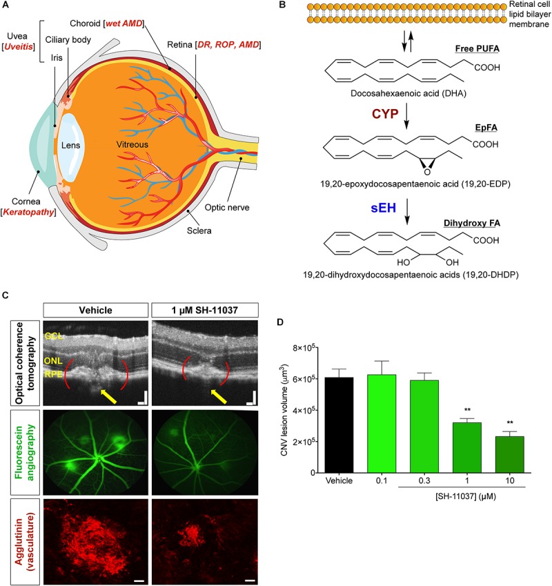
Schematic representation of the eye and PUFA metabolism by the CYP-sEH pathway, and effects of sEH inhibition in vivo. (A) Common eye diseases and associated structures in the human eye. (B) Retina has the highest concentration of ω-3 docosahexaenoic acid of all fatty acids. The regulation of bioactive epoxygenated fatty acids takes place through production by cytochrome P450 epoxygenase and degradation by soluble epoxide hydrolase (sEH). ω-3 fatty acids shown; the same pathway acts on ω-6 fatty acids. (C,D) sEH inhibitor SH-11037 dose-dependently suppresses L-CNV lesion volumes. Figure modified from Sulaiman et al. (2016). (C) Representative imaging data. Optical coherence tomography obtained 7 days post-laser. Yellow arrows highlight regions containing CNV. Scale bars = 100 μm. Fluorescein angiography images 14 days post-laser and confocal microscopy images for agglutinin-stained CNV lesions 14 days post-laser. Scale bars = 50 μm. (D) Quantification of CNV lesion volumes from Z-stack images at day 14 using ImageJ software. ∗∗P < 0.01, one-way ANOVA, Tukey’s post hoc tests, Mean ± SEM, n = 12 eyes/treatment. AMD, age-related macular degeneration; CYP, cytochrome P450 epoxygenase; DR, diabetic retinopathy; EpFA, epoxygenated fatty acid; FA, fatty acid; PUFA, polyunsaturated fatty acid; ROP, retinopathy of prematurity; sEH, soluble epoxide hydrolase.