Figure 4.
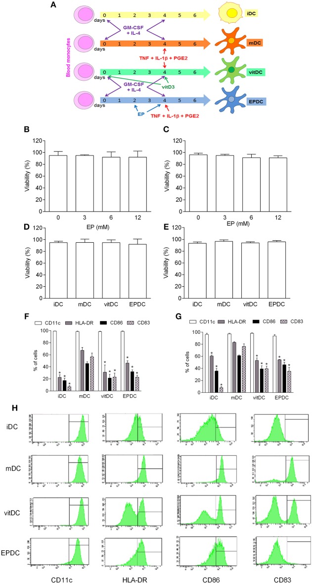
Effects of EP on human DC. MDDC were propagated from peripheral blood monocytes and matured in the presence of TNF+IL-1β+PGE2 (mDC, vitDC, tEPDC) or were left immature without the treatment (iDC) as depicted (A). MDDC were obtained from healthy subjects (B,D,F,H) or from individuals affected by multiple sclerosis (C,E,G). EP was applied in various concentrations (B,C) or in concentration of 6.2 mM (D–H, EPDC). Vitamin D3 (vitDC) was applied in concentration of 1 nM. Viability (B–E) was determined by 7AAD test. Proportion of cells expressing the listed antigens was determined by cytofluorimetry (F–H). Data are presented as mean + SD from at least five individuals. Data in H are representative plots. *p < 0.05 vs. mDC, two-tailed t-test.

