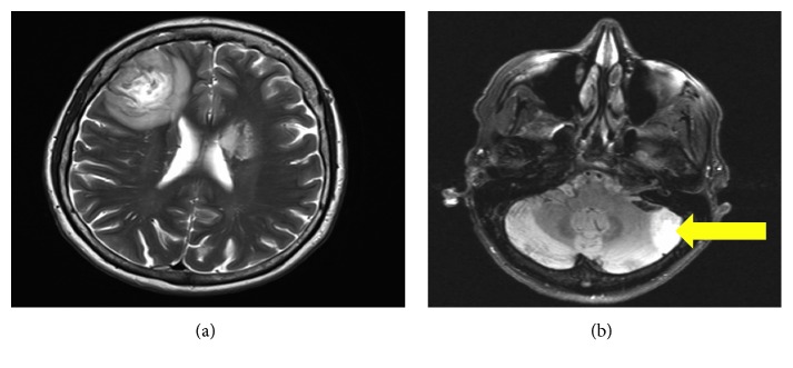
Figure 1.
(a) CT head with contrast showing a 3 cm region of heterogeneous low density in the right frontal lobe. A second, 1.5 to 2 cm region of low attenuation in the anterior aspect of the left basal ganglia consistent with an intraaxial mass lesion compressing the left lateral ventricle. (b) MRI brain with contrast showing a central cavitary, contrast-enhancing lesion involving the lateral left cerebellar hemisphere (see arrow).

