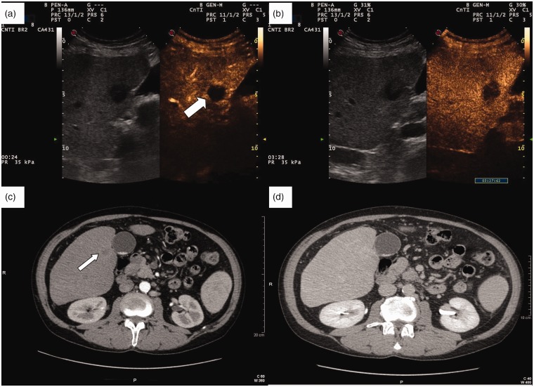
Figure 2.
(a) Contrast-enhanced ultrasound (CEUS) rim-like appearance of a combined hepatocellular–cholangiocellular carcinoma (CHC) nodule during the arterial phase (white thick arrow). (b) The enhancement of the rim fades out in late phase (3 min 26 sec post-injection), with a persistently hypoechoic central area. (c) Contrast-enhanced computed tomography (CT) of the same CHC nodule shows a mild arterial rim-like enhancement (thin arrow). (d) The enhancement progresses towards being mild homogeneous centripetal during the portal-late phase.

