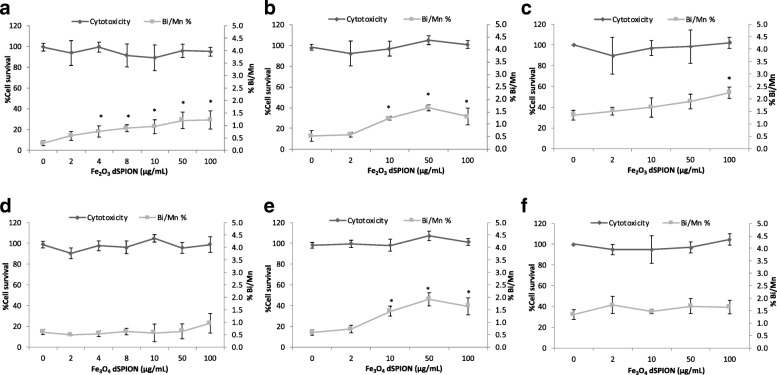
Fig. 4.
Quantification of chromosomal damage and cell viability of 16HBE14o- cells following dSPION exposure. a Mono-cultured 16HBE14o- cells treated with γ-Fe2O3 b 16HBE14o- cells treated with γ-Fe2O3 DTHP-1macrophage conditioned media c 16HBE14o- cells pre-treated with NAC and exposed to γ-Fe2O3 DTHP-1 macrophage conditioned media. d Mono-cultured 16HBE14o- cells treated with Fe3O4 e 16HBE14o- cells treated with Fe3O4 DTHP-1 macrophage conditioned media f 16HBE14o- cells pre-treated with NAC and exposed to Fe3O4 DTHP-1 macrophage conditioned media, *p < 0.05 when compared to negative control (0 μg/ml). For all CBMN assays MMC (0.01 mg/ml) was used as a positive control – (micronuclei fold increase 2.5–2.8%) (n = 3)

