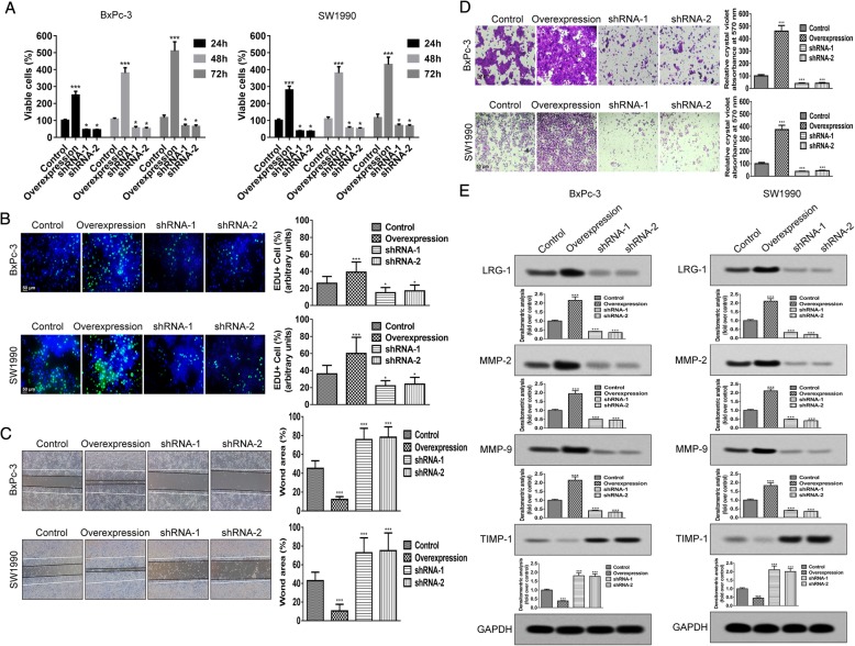
Fig. 2.
LRG-1 promotes PDAC cell viability, proliferation, invasion and migration in vitro. a MTT assays at 24, 48, or 72 h in LRG-1 knockdown and overexpression PDAC cells. b EdU incorporation assay at 48 h in LRG-1 knockdown and overexpression PDAC cells. EdU (green) was used to label proliferating cells and the nucleus was stained with DAPI (blue). Wound-healing assay (c) and Transwell cell invasion assay (d) at 48 h in LRG-1 knockdown and overexpression PDAC cells. e The protein level of MMP-2, MMP-9 and TIMP-1 at day 3 in LRG-1 knockdown and overexpression PDAC cells. Data are presented as the mean ± SD, *p < 0.05; **p < 0.01; ***p < 0.001

