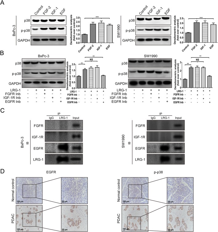
Fig. 5.
The role of EGFR in LRG-1-induced p38 phosphorylation. a Levels of phosphorylated and total p38 were examined by Western blot after 30-min incubation of cells with FGF-2 (20 ng/ml) or IGF-1 (100 ng/ml) or EGF (100 ng/ml). b The expression of phosphorylated and total p38 in both cell lines after 30-min incubation with LRG-1 (500 ng/ml) and FGFR inhibitor (PD173074, 0.2 μM) or IGF-1R inhibitor (OSI-906, 5 μM) or EGFR inhibitor (erlotinib, 5 μM). c Co-immunoprecipitation (Co-IP) between LRG-1 and FGFR or IGF-1R or EGFR. d Immunohistochemistry images of EGFR and p-p38 in human PDAC and normal pancreas tissues. Data are presented as the mean ± SD, NS = not significant; **p < 0.01; ***p < 0.001

