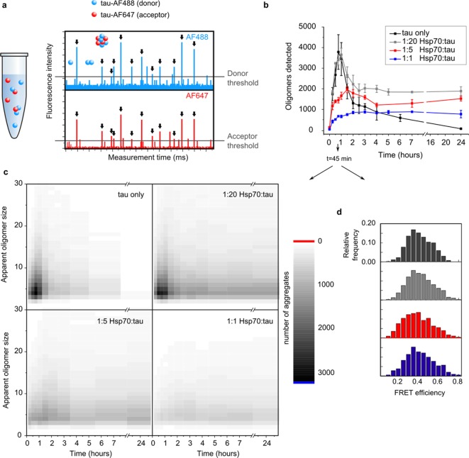
Figure 3.
smFRET shows a reduced number of nuclei in the presence of Hsp70. (a) Typical smFRET spectrum obtained from dual-labeled protein aggregates on a confocal microscope. Single aggregates passing through the confocal volume gave rise to single FRET events which were quantified with regard to frequency, intensity, and FRET efficiency. (b) Evolution of K18 ΔK280 tau oligomers as a function of aggregation time. N = 3, error bars are s. e. m. (c) Apparent sizes of oligomers formed in the absence and presence of Hsp70 after 45 min of aggregation. Sizing reveals a strong decrease in the population of small nuclei formed in the first hour of aggregation. (d) FRET efficiencies of oligomers in the absence and presence of Hsp70 after 45 min of aggregation. Black, tau only; gray, 1:20 Hsp70/tau; red, 1:5 Hsp70/tau; blue, 1:1 Hsp70/tau.

