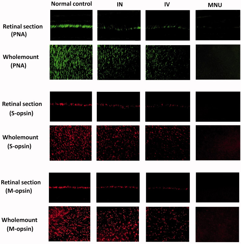
Figure 4.
In the normal control mice, PNA staining was evident at the outer segments of photoreceptors. A pronounced proportion of PNA staining was retained in the retinal sections of the INas administered group. Conversely, no PNA staining was found in the retinal sections of the MNU group. The cone photoreceptors in the dorsal-temporal (DT) and dorsal-nasal (DN) quadrants were preferentially preserved by INas delivery of EPO. The M- and S- opsin positive cells were lost in the retinal section and whole mount of the MNU group. On the other hand, the M- and S-opsin positive cells were evident in the retinal section and whole mount of the INas administered group. The M- and S--opsin staining of the IVen group was prominently weaker than that of the INas administered group. In the INas group, the M- and S--opsin positive cells were distributed throughout the retinal whole mount. However, the distribution was not uniformly equal and formed a topographic gradient across retina: the highest number of M-opsin positive cell was in the DT quadrants and the fewest cell in the ventral-nasal VN quadrant; the highest number of S-cone opsin positive cell was in the VN quadrants and the fewest cell in the DT quadrant.

