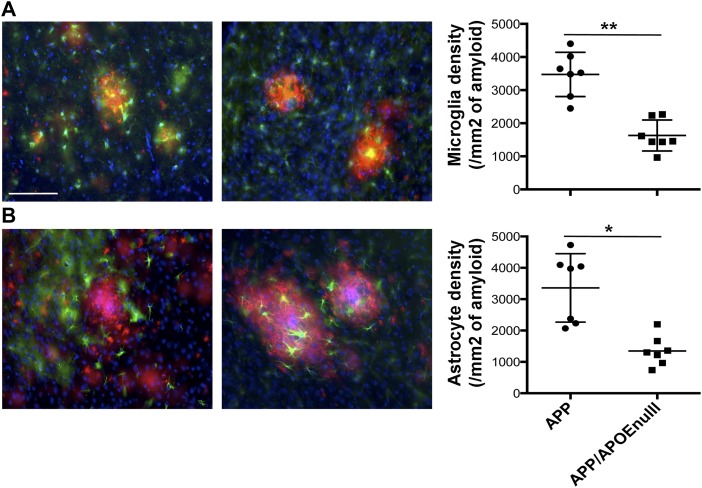
Figure 6. Decreased glial reactivity in APP/PSEN1 mice lacking APOE.
(A) RepreseAPP/PSEN1 and APP/PSEN1/APOEnull adult mice. (B) Representative images (left panel) and stereological evaluation (right panel) of the density of GFAP-positive astrocytes (green) around amyloid deposits (red, after staining using a rabbit anti-Aβ antibody, IBL) in APP/PSEN1 and APP/PSEN1/APOEnull adult mice. In each case, the total number of Iba-1–positive microglia or GFAP-positive astrocytes present in less than 50 μm around a plaque was reported to the surface of the plaque. Scale bar = 100 μm. n = 7 mice/group (10–12 mo “adult” cohort); Mann–Whitney test. *P < 0.005, **P < 0.001.

