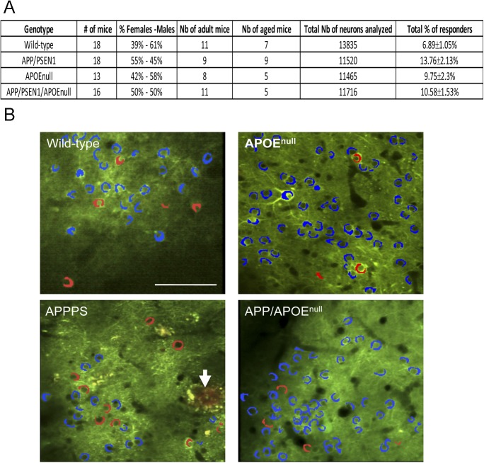
Figure S1. Animal cohort used for recording visually evoked neuronal responses.
(A) Table summarizing the characteristics of the animal cohort used for in vivo recording of visually evoked responses. (B) Representative two-photon images of AAV-CBA-YC3.6 transduced neurons in the visual cortex of wild-type, APP/PSEN1, APOEnull, and APP/PSEN1/APOEnull, indicating the presence of a small proportion of responsive cells (red) as compared with nonresponsive cells (blue). Amyloid deposits were visualized after intraperitoneal injection of Methoxy-XO4 in APP/PSEN1 (arrow) and APP/PSEN1/APOEnull mice. Scale bar = 100 μm.

