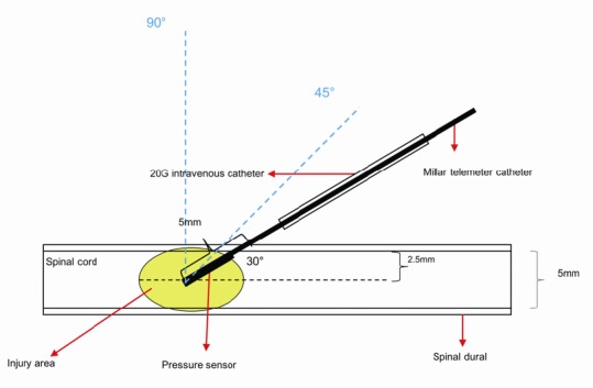
Figure 3.

The pressure probe is inserted into the spinal cord at three different angles.
The spinal cord has a diameter of 5 mm. A 20 G intravenous catheter was used to penetrate the dura with a hole and avoid damaging the blood vessels of the spinal cord. The trocar was then withdrawn, and the pressure probe was inserted along the cannula to the center of the spinal cord. The length of the pressure catheter inside of the spinal dura was 5 mm. The angle of the probe shown to the spinal cord is 30°. Dotted lines indicate angles of 45° and 90°.
