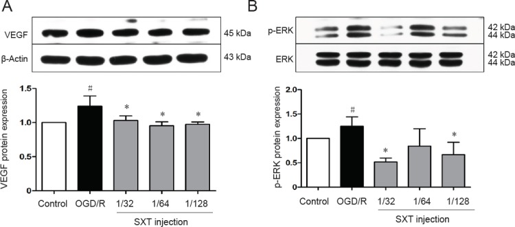
Figure 8.

Effect of SXT on VEGF expression and ERK1/2 phosphorylation in bEnd.3 cells after OGD/R injury.
Cells subjected to 6-hour OGD followed by reperfusion for 18 hours were administered Shuxuetong injection at 1/32, 1/ 64, and 1/128 concentrations (diluted 32-, 64-, and 128-times). Protein expression levels of VEGF (A) and p-ERK1/2 (B) levels were measured by western blot assay. Relative VEGF protein expression was expressed as optical density value relative to the control group after normalizing to β-actin optical density value. Relative p-ERK1/2 protein expression was expressed as optical density value relative to control group after normalizing to ERK1/2 optical density value. Data are expressed as mean ± SD (mean from three independent experiments; one-way analysis of variance followed by least significant difference test). #P < 0.05, vs. control group; *P < 0.05 vs. OGD/R group (n = 3). SXT: Shuxuetong injection; OGD/R: oxygen-glucose deprivation/reperfusion; VEGF: vascular endothelial growth factor; ERK1/2: extracellular signal regulated protein kinase 1/2; p-ERK1/2: phosphorylated extracellular signal regulated protein kinase 1/2.
