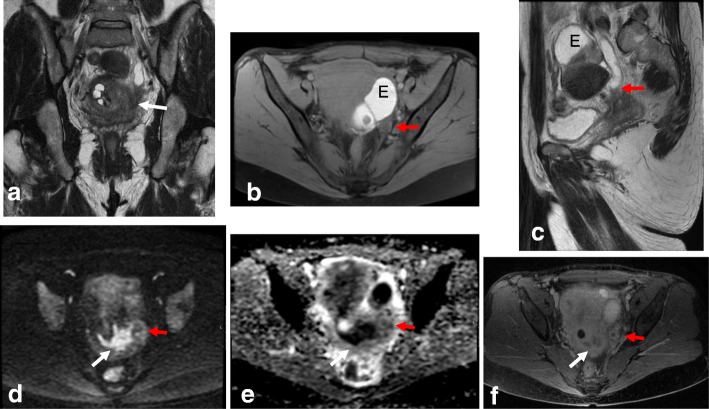
Fig. 16.
MRI reveals FIGO IIIB cervical cancer in a 38-year-old woman with adenomyosis and endometriosis in the left ovary. Visualization by coronal T2WI (a), axial FS T1WI (b), and sagittal T2WI (c) reveals a mass extending to the left parametrium and involving the left ureter (red arrows) below the endometrioma (E), with a subsequent hydronephrosis. d, e DWI (b = 1000/ADC map) reveals diffusion restriction of the cervical mass, with extension to the left ureter. f DCE sequence shows enhancement of the cervical mass and ureter wall

