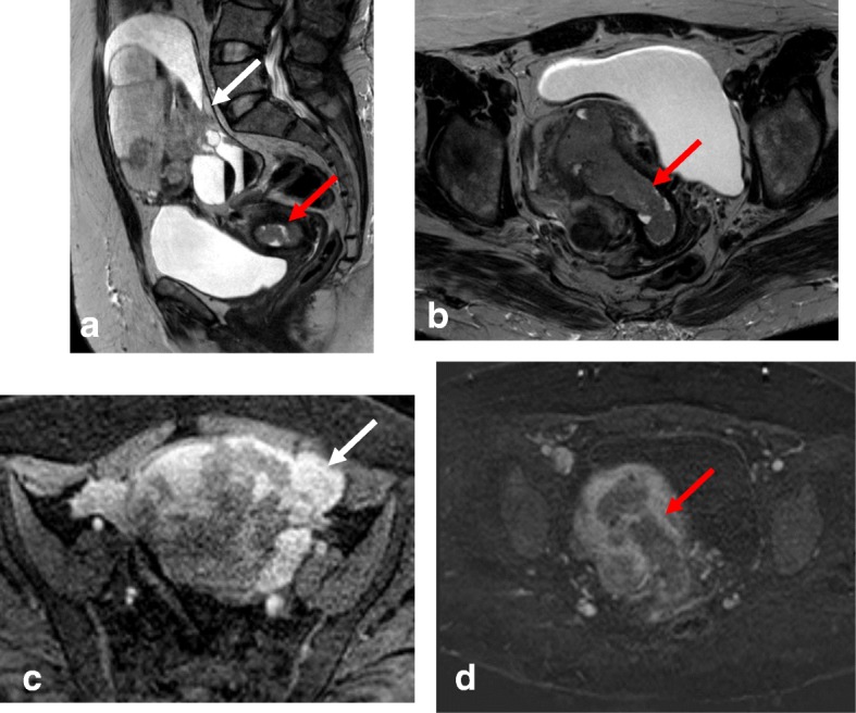
Fig. 8.

Synchronous endometrial and ovarian cancer. Visualization by sagittal T2WI (a) and axial oblique T2WI (b) reveals a large multicystic ovarian mass (white arrow) and endometrial cancer (red arrow). c, d DCE imaging shows heterogeneous uptake in the ovarian mass (white arrow) and hypoenhancement in the endometrial mass (red arrow)
