
Fig. 13.
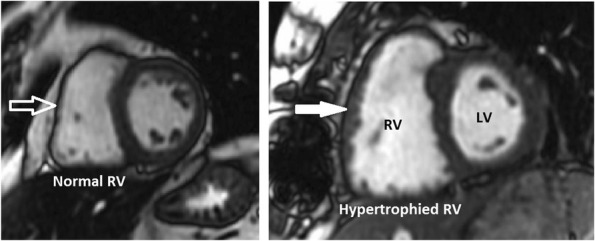
Right ventricular wall hypertrophy. Short axis SSFP MRI images demonstrate normal (open white arrow) and hypertrophied (closed white arrow) RV wall
Official websites use .gov
A
.gov website belongs to an official
government organization in the United States.
Secure .gov websites use HTTPS
A lock (
) or https:// means you've safely
connected to the .gov website. Share sensitive
information only on official, secure websites.
Right ventricular wall hypertrophy. Short axis SSFP MRI images demonstrate normal (open white arrow) and hypertrophied (closed white arrow) RV wall