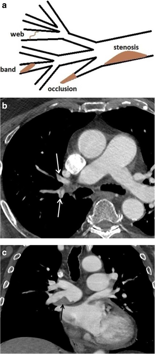
Fig. 20.

Configurations in CTEPH. a Figure demonstrating findings of CTEPH including stenosis, web, band, and occlusion. b CTA axial image of web/band (white arrows). c CTA axial image demonstrates eccentric thrombus narrowing the lumen of the right main pulmonary artery (black arrow)
