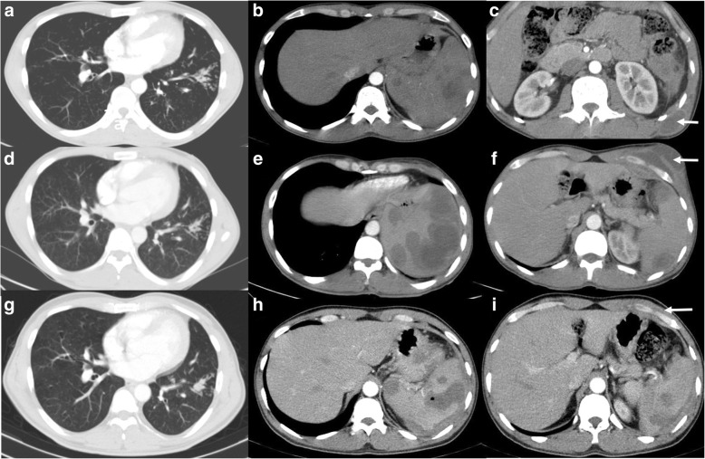
Fig. 2.
Chronic melioidosis with TB. Forty-one-year-old gentleman, newly diagnosed to have diabetes mellitus, presented with respiratory symptoms, (a) CT thorax showed the tree in bud opacities in the left lower lobe. Upper abdomen sections reveal (b) multiple hypodense foci in the spleen suggestive of small abscesses and (c) a small abscess in the left posterior lower chest wall (arrow). Cytology from the chest wall showed necrotising granulomatous inflammation, and antituberculous treatment was started. Two years later, he presented with new onset of left anterior chest wall swelling, for which CT thorax and abdomen were performed which showed (d) mild reduction in the ‘tree in bud’ opacities, but (e) increase in the size and number of splenic abscesses and (f) interval development of the left anterior lower chest wall abscess (arrow). Pus culture from the chest wall grew Burkholderia pseudomallei. Follow-up CT after 6 months showed, (g) stable tree in bud opacities in the lung, (h) near complete resolution of the chest wall abscess (arrow) but with the presence of perisplenic fluid with persisting splenic lesions (h and i), suggestive of contained rupture

