Fig. 8.
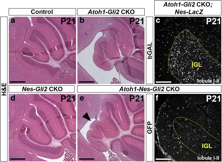
NEP-derived cells failed to populate the IGL at P21. a, b, d and e H&E staining of sagittal sections of anterior cerebellar vermis of P21 Gli2flox/flox (Control, a), Atoh1-Cre/+; Gli2flox/flox (Atoh1-Gli2 CKO, b), Nes-FlpoER/+; R26MASTR/+; Gli2flox/flox (Nes-Gli2 CKO, d) and Atoh1-Cre/+; Nes-FlpoER/+; R26MASTR/+; Gli2flox/flox (Atoh1-Nes-Gli2 CKO, e) mice injected with Tm at P0. Note that inactivation of Gli2 in Nestin-expressing cells inhibits the compensation mechanism in the anterior vermis (black arrowhead in e). c and f FIHC detection of the indicated proteins on mid-sagittal cerebellar sections (lobule I-II) of P21 Atoh1-Cre/+; Gli2flox/flox; Nes-FlpoER/+; R26FSF-LacZ/+ (Atoh1-Gli2 CKO; Nes-LacZ, c) and Atoh1-Cre/+; Nes-FlpoER/+; R26MASTR/+; Gli2flox/flox (Atoh1-Nes-Gli2 CKO, f) mice injected with Tm at P0. IGL is indicated by the yellow doted line. Scale bars represent 500 μm (a, b, d and e) and 100 μm (c and f)

