Fig. 2.

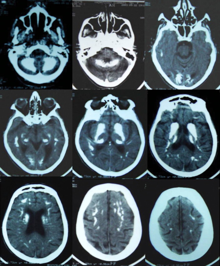
Series of brain CT scanning showing widespread, symmetrically located calcifications in both the frontal lobes, subcortical nuclei, paraventricular region, brain fornix, as well as both cerebellar hemispheres.

Series of brain CT scanning showing widespread, symmetrically located calcifications in both the frontal lobes, subcortical nuclei, paraventricular region, brain fornix, as well as both cerebellar hemispheres.