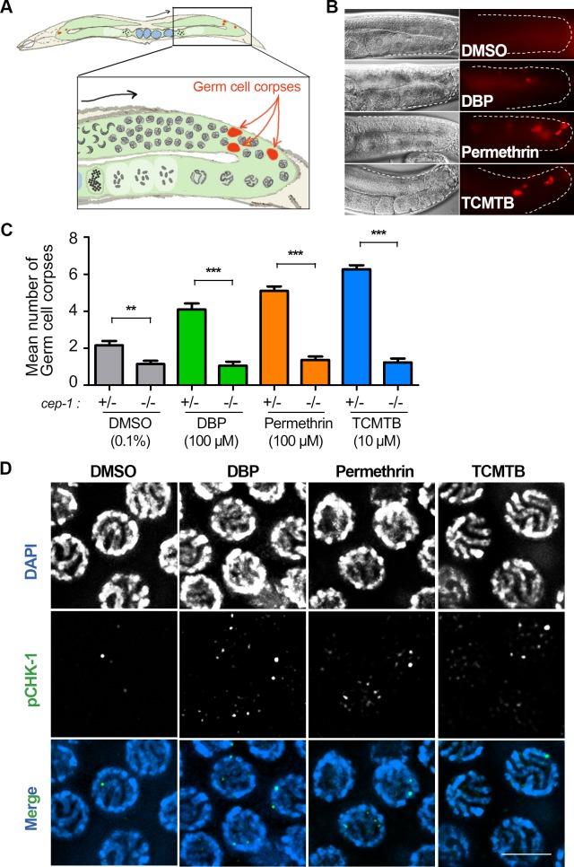
Fig 3. DBP, Permethrin, and TCMTB exposures lead to p53/CEP-1-dependent increased germ cell apoptosis and CHK-1 activation.
(A) Schematic representation of C. elegans and the area where DNA damage checkpoint activation of germ cell apoptosis is detected in the germline. Inset represents zoom-in of one gonad arm. Black arrow indicates orientation of progression through meiosis while red arrows indicate germ cell corpses (red) observed at late pachytene near the gonad bend. (B) Chemical exposures caused a significant increase in the number of germ cell corpses observed at late pachytene compared to vehicle alone. Gonads are traced to facilitate visualization. On the left is the Nomarski optics view and to the right are the acridine orange stained germ cell corpses (red). (C) Graphical representation showing mean number of germ cell corpses detected for each indicated chemical. Levels of germ cell corpses were significantly reduced in a p53/cep-1-dependent manner. Note that basal level of toxicity for DMSO, as previously described [42,103], is also reduced in a cep-1 mutant background. Analysis was done for three independent biological repeats. More than 30 gonads were scored for each chemical. Error bars represent SEM. **P<0.01, ***P<0.0001 by the two-tailed Mann-Whitney test, 95% C.I. (D) High-resolution images of mid to late pachytene nuclei from whole-mounted gonads immunostained for phospho CHK-1 (pCHK-1; green) and co-stained with DAPI (blue). Elevated levels of pCHK-1 were observed in chemical-treated worms compared to control. Scale bar, 5 μm.

