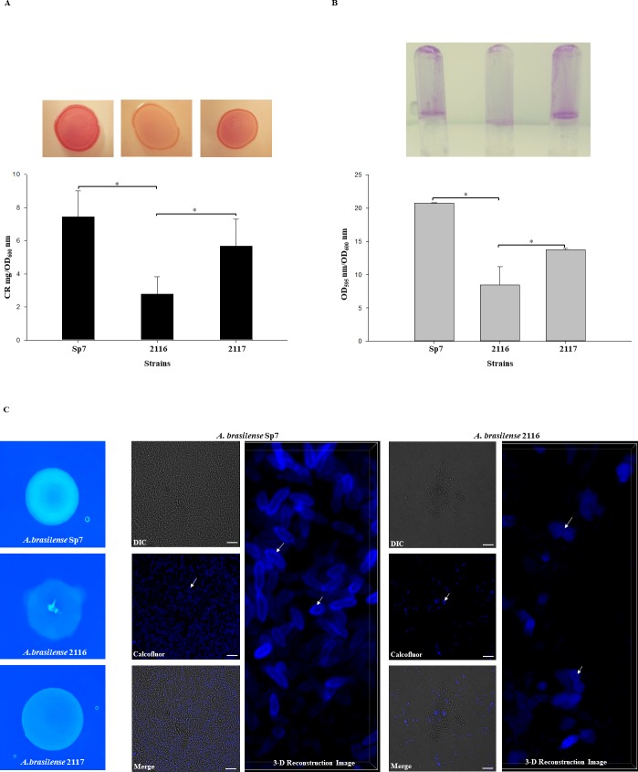
Fig 6. Biofilms and EPS production by the A. brasilense Sp7, 2116, and 2117 strains.
(A) CR staining assay. Cells were grown on agar-solidified CR medium inoculated with 10 μL of 1.0 OD600 nm of each culture strains for 72 h at 30 ºC. For EPS quantification, the strains were cultured as described in Material and Methods and incubated at 30°C for 5 days. CR binding was expressed as mg CR/OD600 nm. (B) For biofilm production cells were grown statically for 5 days at 30 ºC on NFb* with 27mM malate as a C source + 10mM D-alanine. Biofilm formation was visualized by Crystal violet staining as well quantified and normalized per OD600 nm of growth. All data are the average from three independent experiments performed in duplicate. Error bars indicate standard errors of the means as compared with values observed for WT. For all data, P was < 0.001 as assessed by Student´s t-test. (C) Calcofluor white colorant M2R (CWC) staining. Cells were grown on agar-solidified NFb* supplemented with CWC and inoculated with 10 μL of 1.0 OD600 nm of each culture strains for five days at 30 ºC. The cell fluorescence was observed under was observed with a Nikon Eclipse Ti-E C2+. Bars represent 10 μm. The white arrows indicate EPS.

