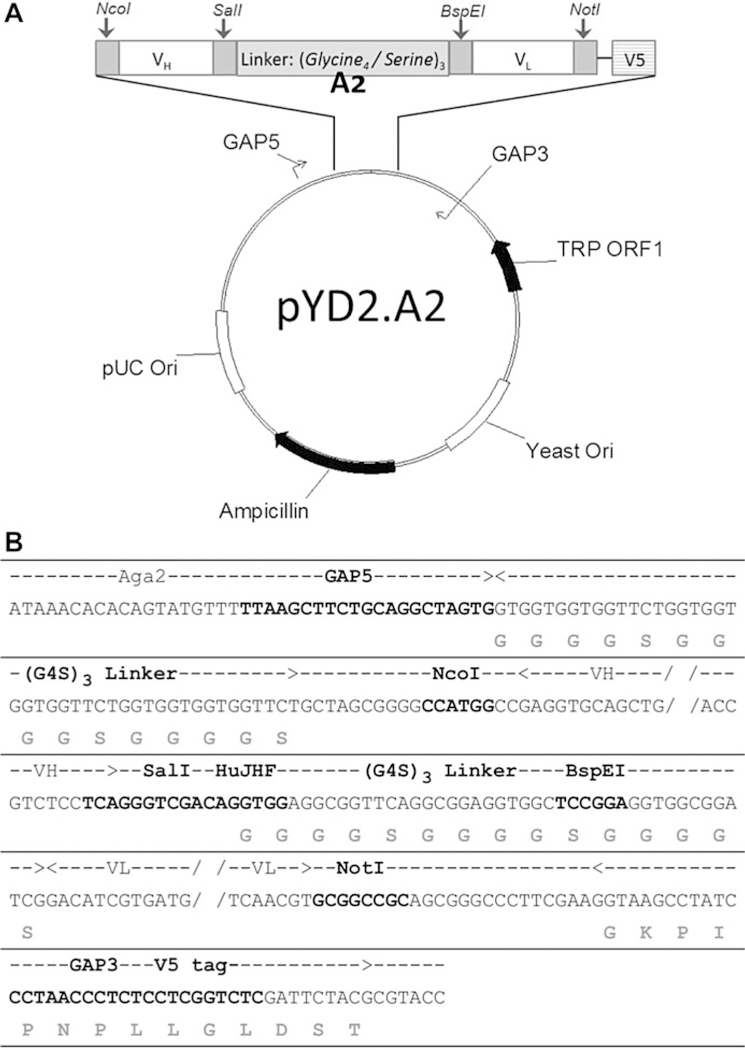
Fig. 1. Vector map and sequence of pYD2.A2 plasmid.

(a) Vector map of pYD2.A2 plasmid showing the relative position of genes, linkers, primer sites and restriction digestion sites. (b) Sequence showing the insert of A2 region in the vector. The bold nucleotides in the middle row indicate 1) GAP5, GAP3 and HuJHF primer binding sites and 2) the restriction digestion sites for NcoI, SalI, BspEI and NotI. The grey amino acids on the bottom row indicate the (G4S)3 linkers and V5 tag. The dashed line with arrows on the top row indicate the span of the (G4S)3 linkers, VH insert, VL insert and V5 tag
