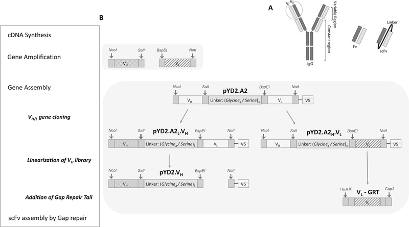
Fig. 2. Illustration of method 3.1 section: Construction of yeast display library.

(a) Schematic representation of a scFv fragment. During library construction, VH and VL are randomly paired via a flexible linker to form scFvs. The quality of a library construction is dependent on the degree of scFv pairing diversity. (b) Schematic overview of scFv construction is boxed on the left, with a list of major steps involved in scFv construction. The corresponding diagram on the right illustrates the designated products derived from PCR, restriction digestion and ligation reactions. Vector pYD2.A2 displaying A2-scFv was created by modifying the pYD2 vector, which comprises a (Gly4/Ser)3 linker region carrying SalI and BspEI restriction sites, and NcoI and NotI restriction sites flanking the inserted A2-scFv. An in-frame V5 epitope allows detection of scFv fusion product and normalization of scFv surface expression through immunofluorescence labeling of V5-tag. Patterned filled bars represent amplified VH and VL from all of the heavy and light chain family genes using partially degenerated primers
