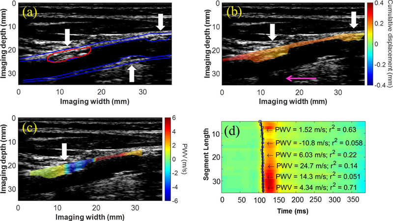
Figure 2:

(a) B-Mode from the right CCA of a 72-y.o. female (50–79% stenosis) containing atherosclerotic lesions on both the anterior and posterior walls (white arrows). (b) Decreased cumulative displacement was observed in the anterior wall plaques (white arrows). The presence of the acoustic shadow (pink arrow) obstructing a portion of the posterior wall suggests that the plaque directly above is moderately calcified. (c) The PWV map reveals a region where the wave appears to converge inside the plaque (white arrow). (d) Piecewise PWV measurements overlaid on the spatiotemporal map.
