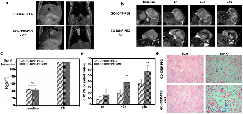
Figure 3.
In vivo MR imaging (n = 5). (a) T2-weighted MR images of the mouse liver. The images were taken 24 h after intravenous injection of GO-IONP-PEG, with or without an external magnetic field (MF). (b) T2-weighted MR images of tumors. Relative to the prescans, signal reduction was observed in tumors after GO-IONP-PEG injection, and the reduction level was gradually elevated. When there was a magnet attached to the tumor (bottom), the signal reduction was enhanced. (c) Quantitative analysis of R2 in the liver before and 24 h after the particle injection, based on the imaging results from panel a. ns: no statistical difference. (d) Quantitative analysis of relative R2 change (dR2) at 6, 12, and 24 post injection, based on the imaging results from (b). Significant R2 increase was observed when an external magnetic field was applied (*, p < 0.05; **, p < 0.01). (e) Prussian blue staining with liver and tumor samples. Although the liver uptake was comparable, using a magnet led to enhanced particle accumulation in the tumor.

