
Figure 2. Compilation of all Gammaproteobacteria flagellar motors imaged to date by ECT.
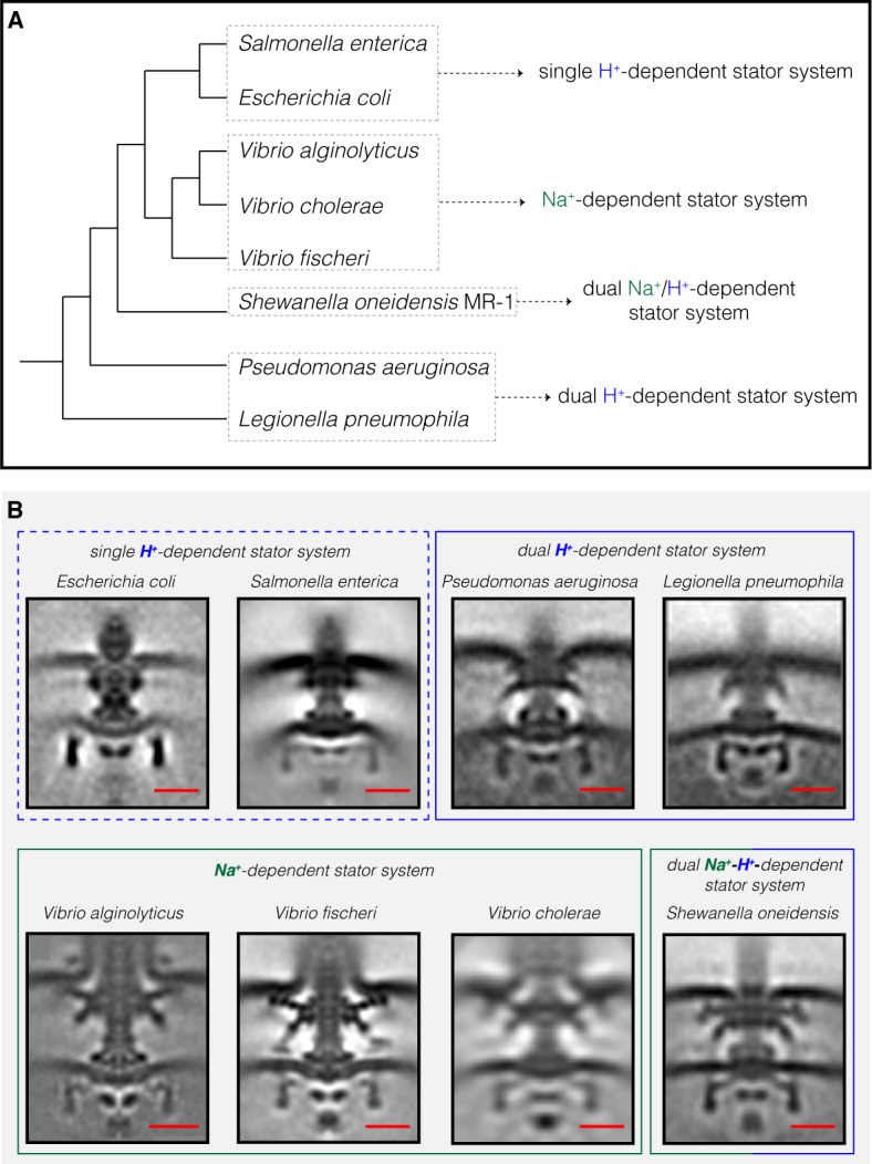
(A) A phylogenetic tree of the eight Gammaproteobacteria species with available ECT structures of their flagellar motors. This tree was made based on (Williams et al., 2010). (B) Central slices of sub-tomogram averages are shown for the eight Gammaproteobacteria flagellar motors revealed by ECT, including the three structures solved in this study (P. aeruginosa, L. pneumophila and S. oneidensis). The motors are classified based on their stator system: single H+-driven (dashed blue box), dual H+-driven (blue box), Na+-driven (green box) or dual Na+-H+-driven (green-blue box). E. coli EMDB 5311, S. enterica EMDB 3154, V. fischeri EMDB 3155, V. cholerae EMDB 5308, V. alginolyticus is adapted from Zhu et al., 2017. Scale bars are 20 nm.