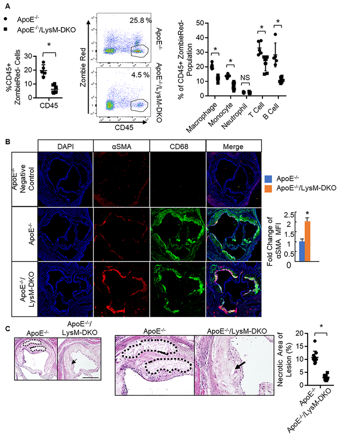
Figure 2. Myeloid-specific deletion of epsins decreases immune and inflammatory cell content in the aorta, increases smooth muscle cell content, and decreases necrotic core content within the atheroma.
A. Aortas from ApoE−/−(n=5) and ApoE−/−/LysM-DKO (n=5) fed Western diet (WD) were isolated, digested, cells isolated, labeled with CD45 (hematopoietic cells), CD11b (granulocytes, monocytes, macrophages, dendritic cells, NK cells), F4/80 (macrophage; F4/80+CD11b+ defined as macrophage), CD19 (B cells), TCRβ (T cells), Ly6C (monocytes), Ly6G (neutrophils defined as Ly6C+;Ly6G+), and analyzed via flow cytometry. *ApoE−/−/LysM-DKO group vs. ApoE−/− group, P<0.01. Aortic root sections from ApoE−/− (n=8) and ApoE−/−/LysM-DKO (n=10) fed WD for 20 weeks (B) and 25 weeks (C) were stained with CD68 (macrophages, green) and α-Smooth Muscle Actin (αSMA) (red, smooth muscle cells) in B and H&E in C. Scale bar=200μM. *ApoE−/−/LysM-DKO group vs. ApoE−/− group, P<0.01.

