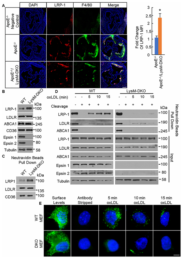
Figure 4. LRP-1 total and surface levels are increased with decreased internalization in plaques and macrophages from myeloid-specific epsins deficient mice.
A. Aortic root sections from ApoE−/− (n=8) and ApoE−/−/LysM-DKO (n=8) mice fed Western diet (WD) for 14 weeks were stained with LRP-1 (red) and F4/80 (Mϕ, green). Scale bar=200μM. *ApoE−/−/LysM-DKO group vs. ApoE−/−, group, P<0.01. B. Elicited peritoneal Mϕs from WT (n=5) and LysM-DKO mice (n=5) were lysed forwestern blot (WB). C. Elicited peritoneal Mϕs from WT (n=3) and LysM-DKO (n=3) mice were biotinylated and processed for neutravidin beads pull down followed by WB. D. Elicited peritoneal Mϕs from WT (n=5) and LysM-DKO (n=5) mice were biotinylated, treated with or without oxLDL (10μg/mL) for indicated time points, and treated with or without cleavage buffer followed by processing for neutravidin beads pull down and WB. E. WT and DKO MEFs transfected with FLAG-tagged LRP1 were labeled with anti-FLAG antibodies. Cells were treated with oxLDL (10μg/mL) for the indicated time points followed by treatment with antibody stripping buffer and staining with secondary antibodies and DAPI. Images were taken at 60×. Scale bar=5μm.

