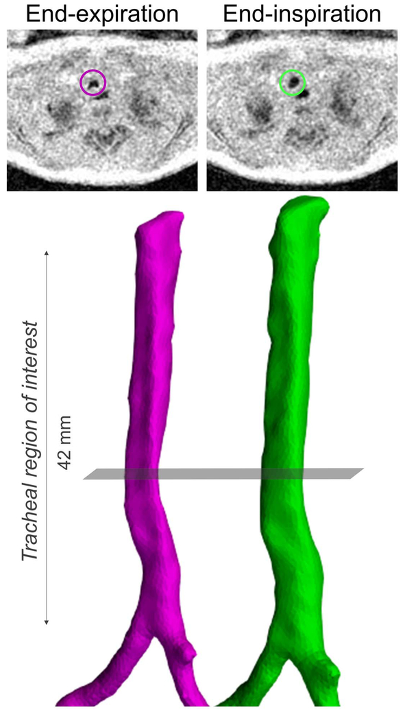
Figure 2.
Tracheal surface renderings created from segmentations from retrospective respiratory-gated ultrashort echo-time (UTE) MRI (pink - end-expiration; green - end-inspiration) in a neonatal subject with congenital diaphragmatic hernia (CDH). Axial MR images shown above each airway rendering correspond to the same part of the respiratory cycle (end-expiration or end-inspiration) and are at the level of the grey plane. In both the MR images and airway renderings, the regional collapse of the trachea occurring at endexpirationis clearly visible.

