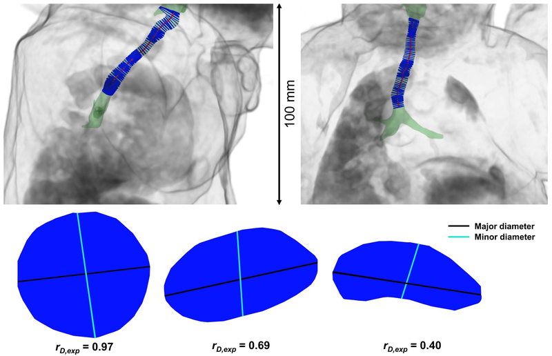
Figure 3.
A neonatal airway surface rendering (green) shown within a body volume rendering (grey) from a neonatal ultrashort echo-time (UTE) MRI scan, retrospectively respiratory-gated to end-inspiration. A center-line is defined along the airway (red), with a series of luminal cross-sections (blue) bounded by the airway and orthogonal to the center-line. Sagittal and coronal views are shown at left and right, respectively. Note that this subject’s trachea is not aligned with any of the imaging planes, demonstrating the need to define a tracheal center-line for accurate cross-sections of the airway lumen. Three example luminal cross-sections are shown below, demonstrating three different values of rD,exp, with descending values from left to right. The major diameters are shown in black, and the minor diameters are shown in cyan.

