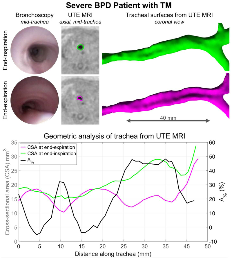
Figure 4.
Representative MRI results for a neonatal patient with severe bronchopulmonary dysplasia (BPD) and bronchoscopically-diagnosed tracheomalacia (TM). Collapse of the posterior tracheal wall at endexpiration is clear on both clinically-obtained bronchoscopic images and retrospectively respiratory-gated ultrashort echo-time (UTE) MRI results. The tracheal lumens are outlined on end-expiration and endinspiration UTE axial images in pink and green, respectively. Gated UTE-based tracheal surfaces are shown, along with corresponding geometric analysis along the intrathoracic trachea: cross-sectional areas (CSA) at end-expiration (pink) and end-inspiration (green); and percentage reduction in CSA (A%, black). Quantitative MRI results of dynamic collapse, particularly notable throughout the middle and lower tracheal regions, agree with qualitative bronchoscopic results.

