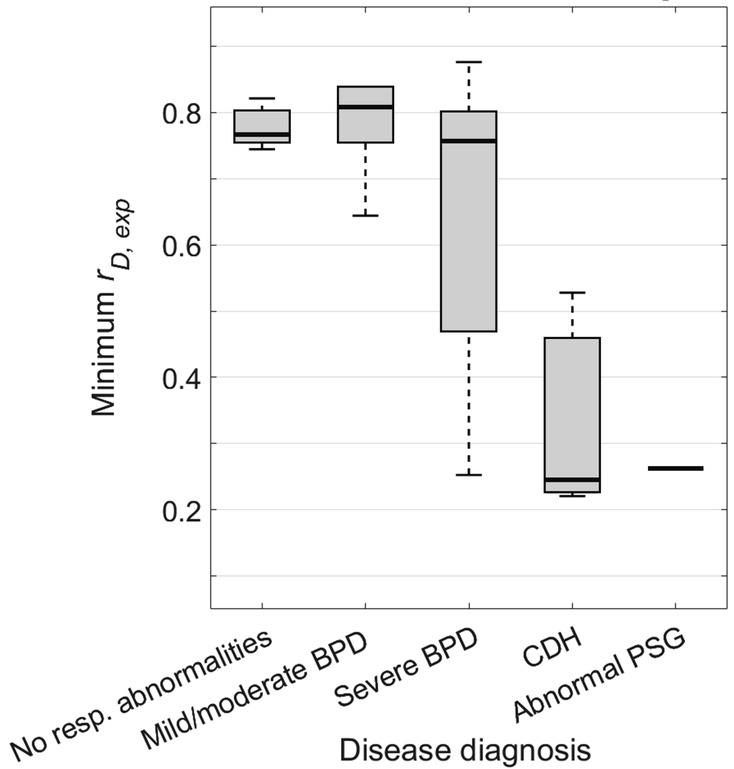
Figure 7.
Comparison of minimum ratio of tracheal minor-to-major diameter at end-expiration (rD,exp) to subjects’ respiratory morbidity group. This measure did not statistically distinguish between subjects with mild/moderate BPD and control subjects (P = 0.859). Subjects with severe BPD have a larger range of minimum rD,exp than control subjects with no respiratory abnormalities and subjects with mild/moderate BPD, though without statistically significant separation (P = 0.138 and 0.132), suggesting that airway disease may be comorbid with lung disease in some but not all severe BPD patients. However, there is a statistically significant difference between the severe BPD group and a combined control and mild/moderate BPD group (P = 0.036). Subjects with CDH also have a lower minimum rD,exp value that is significantly different from control subjects (P < 0.001). Plot elements are represented as follows: median (black line), interquartile range (grey box), and data within 1.5 times the interquartile range below 25% and 75% (black whiskers).

