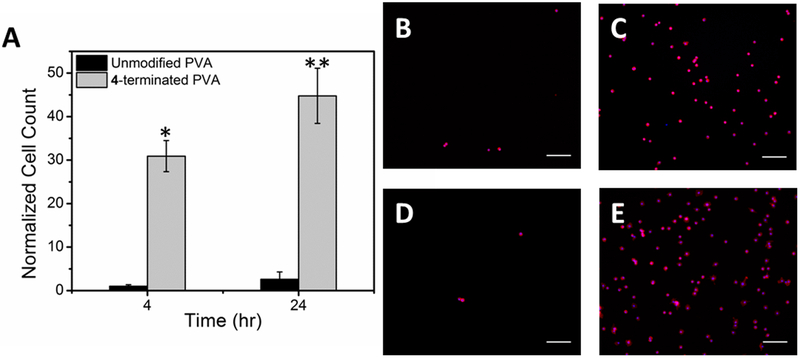
Figure 6.
Primary mature bovine chondrocytes on 4-terminated and unmodified PVA thin film surfaces. (A) Normalized cell count, all data are normalized to the 4 hr count on unmodified PVA. Data are reported as average ± 1 S.E. (n ≥ 16). Images, 20x, of chondrocytes stained with rhodamine phalloidin (red) and DAPI (blue) on (B) unmodified PVA after 4 hr, (C) 4-terminated PVA after 4 hr, (D) unmodified PVA after 24 hr, and (E) 4-modified PVA after 24 hr.(*) denotes statistical difference from the 4 hr control and (**) denotes statistical difference from the 24 hr control; scale bars = 100 μm.

