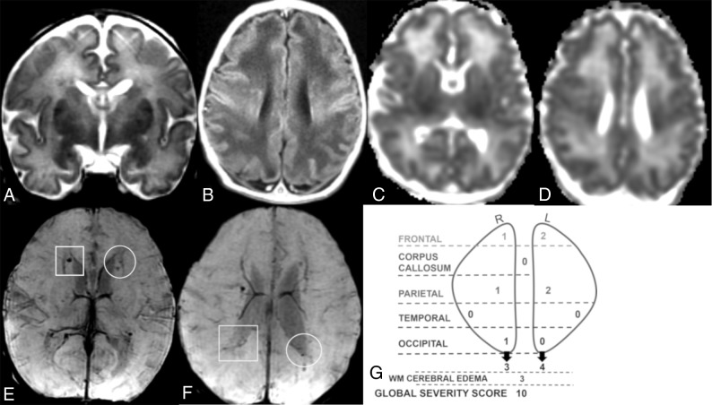
Fig 2.
Coronal T2 (A), axial T1 (B), ADC (C and D), and SWI (E and F) MR images of a 7-day-old girl. Global severity score = 10 (second quartile) (G). There is diffuse bilateral WM cerebral edema (3 points) (A and B) and multiple acute bilateral, asymmetric (left > right) hemorrhagic WM lesions (T1 bright and T2 dark) without restricted diffusion (C–F). On the right, there are mild punctate lesions in the frontal (1 point), parietal (1 point), and occipital WM (1 point, not shown) (E and F, boxes), and on the left, there are moderate punctate lesions in the frontal (2 points) and parietal WM (2 points) (E and F, circles). No corpus callosum or temporal region WM lesions are visible. R indicates right; L, left.

