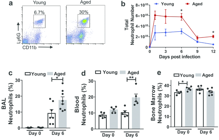
Figure 1. Aging is associated with increased neutrophils within the lung during influenza infection.
Aged (18–22 months of age) and young (2–4 months of age) C57BL/6 mice were intranasally inoculated with PR8 strain influenza virus. Lung tissue was harvested, digested and cells were obtained and suspended to permit incubation with fluorescent monoclonal antibodies followed by flow cytometric analysis.
a Representative flow cytometric plot at day 6 p.i. (post-infection) of a young and aged mouse showing increased proportion of neutrophils (CD11b+, Ly6Ghi) within the lung in the aged mouse than the young mouse. The flow cytometric plots are gated on CD11b+Ly6Ghi cells.
b Absolute number of neutrophils before and during influenza infection in young and aged mice in entire lung (airspace, vasculature and lung interstitium). * P < 0.05, (Mann-Whitney test). n = 3–6 / group/ time point. Data representative of one of two independent experiments. c,d,e Proportion of neutrophils analyzed by flow cytometry in non-infected and infected (day +6 p.i.) young and aged mice in BAL (c) blood (d) and bone marrow (e). * P < 0.05, ** P < 0.01 (Mann-Whitney test). Data representative of one of two independent experiments,which yielded similar results.

