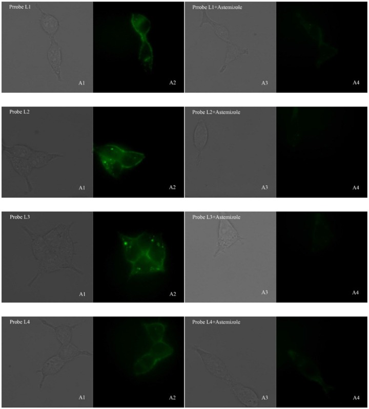
Figure 3.
Fluorescence microscopy imaging of hERG transfected HEK293 cells incubated with 5 μM probe L1, 5 μM probe L2, 1 μM probe L3, 5 μM probe L4 (A1, bright field; A2, GFP channel), respectively. The imaging of inhibition of the hERG channels was accomplished by incubating astemizole (50, 50, 10, 50 μM) with probe L1 (5 μM), L2 (5 μM), L3 (1 μM), and L4 (5 μM) (A3, bright field; A4, GFP channel). All cells were incubated with each probe at 37°C for 10 min and washed immediately. The background was adjusted by ImageJ software. Imaging was performed using a Zeiss Axio Observer A1 microscope with a 63 × objective lens.  .
.

