Figure 4.
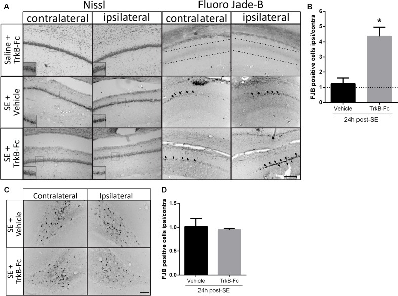
TrkB-Fc increases neuronal death after SE in vivo. (A) Micrographs of hippocampal CA1 stained with Nissl (left panels) or with Fluoro-Jade B (FJB; right panels) show representative brain sections of animals with each treatment. Insets show, at higher magnification, morphological alterations evidenced by Nissl staining. Either TrkB-Fc or vehicle were infused in the hippocampi of one hemisphere (ipsilateral) of control (saline) and SE animals. (B) Quantification of the ipsilateral/contralateral FJB positive neurons in CA1 area. (C) Micrographs of hippocampal hilus stained with FJB show representative brain sections of animals with each treatment. (D) Quantification of the ipsilateral/contralateral FJB positive neurons in hippocampal hilus. Results are expressed as a mean ± SEM of three animals per treatment. Asterisk indicates p < 0.05 compared to vehicle by nested ANOVA. Scale bar = 200 μm.

