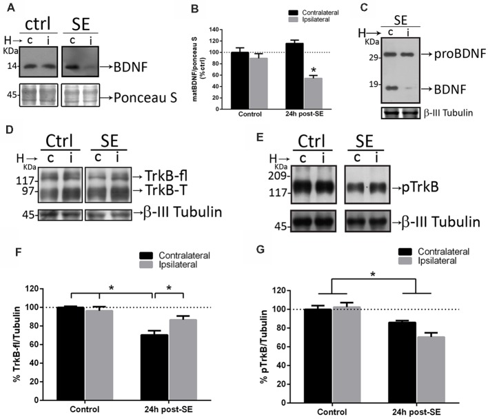
Figure 5.
Sequestering BDNF prevents TrkB signaling. TrkB-Fc was infused in the hippocampi of one hemisphere (ipsilateral, i) of control and SE animals. The dorsal hippocampus from non-infused side (c) and infused side (i) were homogenized separately and analyzed by western blot to determine BDNF (A), proBDNF (C), TrkB (D) and pTrkB (E). Panels (B,F,G) show the quantifications for BDNF (B), TrkB (F) and pTrkB (G). Black bars represent non-infused hippocampus and gray bars TrkB-Fc infused sides. Results are expressed as mean ± SEM of four animals per treatment. Asterisks indicate p < 0.05 by two-way ANOVA followed by Tukey’s post hoc analysis.

